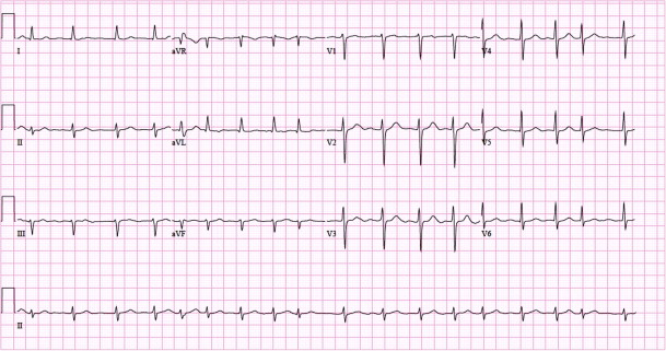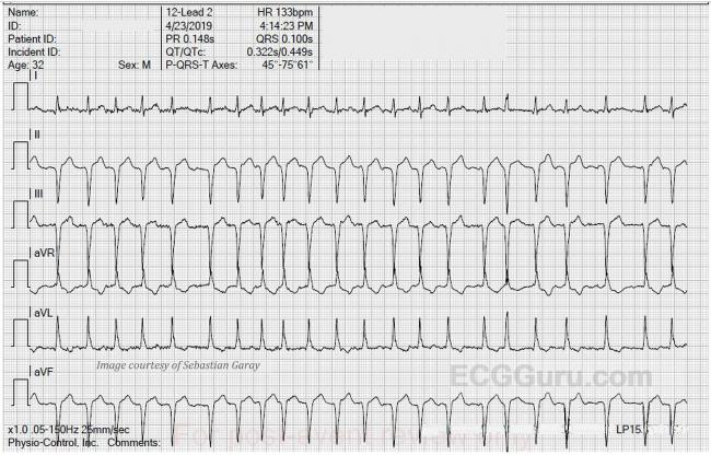
Atrial Fibrillation Ecg Interpretation. Ecg features of atrial fibrillation in wpw are. No p waves a irregular baseline is seen. Rapid ventricular rates may result in degeneration to vt or vf. Atrial fibrillation can occur in up to 20 of patients with wolff parkinson white syndrome wpw the accessory pathway allows for rapid conduction directly to the ventricles bypassing the av node.

Ecg features of atrial fibrillation in wpw are. Atrial fibrillation with bradycardia ecg example 1 atrial fibrillation with bradycardia ecg example 2 atrial fibrillation with bradycardia ecg example 3. No p waves a irregular baseline is seen. Impulses are regulated by the atrioventricular av node which mainly controls the number of impulses that passes along the ventricles. Atrial fibrillation is the rapid firing of impulses in the right atrium with about 350 to 650 beats or impulses per minute. Atrial rate usually 400 to 600 bpm.
No p waves a irregular baseline is seen.
Atrial fibrillation is the rapid firing of impulses in the right atrium with about 350 to 650 beats or impulses per minute. The characteristic ecg changes seen in atrial fibrillation are. Ventricular rate is variable. Atrial fibrillation with bradycardia ecg example 1 atrial fibrillation with bradycardia ecg example 2 atrial fibrillation with bradycardia ecg example 3. Atrial rate usually 400 to 600 bpm. Rapid ventricular rates may result in degeneration to vt or vf.